What is MRI
Magnetic resonance imaging (MRI) is a test that uses a magnetic field, pulses of radio wave energy and a computer system to provide pictures of organs and structures inside the body. In many cases, an MRI provides information that cannot be obtained from an X-ray, ultrasound or CT scan.
For an MRI test, the area of the body being studied is positioned inside a strong magnetic field. The MRI can detect changes in the normal structure and characteristics of organs or other tissues. These changes may indicate diseases caused by trauma, infection, inflammation or tumors. Information from an MRI scan is saved and stored on a computer for further study by a physician.
In some cases a contrast material may be used during the MRI scan to enhance the pictures of certain structures. The contrast material may help evaluate blood flow, detect some types of tumors, and locate areas of inflammation.
Our Equipment
Exeter Hospital’s main MRI unit is a 1.5 Tesla high field Siemens Avanto MRI machine (seen above). The MRI table moves into a short tube that is open at both ends. The well-lit room is spacious with a skylight and windows. Blankets are provided if needed. Music is provided or you may bring a CD/MP3. A ball is given to the patient as a form of communication to the technologist. The technologist can see you at all times, but if you need to stop the exam, all you have to do is squeeze the ball.
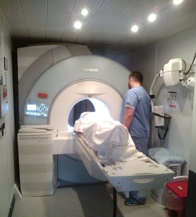
In addition to our fixed unit, Exeter Hospital also offers a mobile 1.5T Siemens Espree Open MRI which is ideal for claustrophobic and larger patients. Please speak to your physician about your options if you feel this is necessary.
Contact Information
Exeter Hospital
5 Alumni Drive
Exeter, NH 03833
Exeter Hospital MRI Department Phone: (603) 580-6244
Exeter Hospital MRI Department Fax: (603) 580-7458
Exeter Hospital Scheduling Department Phone: (603) 580-6966
Exeter Hospital Scheduling Department Fax: (603) 580-6919
What to Expect
You will be asked to lie down on the MRI table. Depending on the exam, you may be positioned head-first or feet-first and your exam will usually take between 30 and 60 minutes. The table is moved into position within the large donut-shaped magnet. The part of the body being imaged needs to be positioned in the center of the magnet. The technologist will be in contact with you at all times visually and through the headphones. You will hear a series of repetitive pulsing or knocking noises. The noise occurs when the images are being taken so it is particularly important to hold very still. Motion causes the images to blur and will extend the exam time since it would be necessary to repeat that sequence. Headphones with music or earplugs are offered to muffle the loud noises heard during the exam.
Your doctor or the radiologist will determine whether an injection of contrast (dye) is necessary to complete your test. The contrast material may help evaluate blood flow, detect some types of tumors, and locate areas of inflammation. This contrast does not contain iodine.
Dietary Restrictions
If you are scheduled for an exam of your bile ducts or liver, we ask that you do not eat or drink anything 4-6 hours prior to your exam.
Registration and Screening
We ask that you arrive to register for your exam 30 minutes prior to your exam’s scheduled time. This will allow you time to go through the registration process and fill out necessary paperwork. After this, you will be brought to the MRI waiting room where we will go over your paperwork to ensure that you are safe to have an MRI. Then, if necessary, you will change into tops and/or bottoms.
For Your Safety
Please wear clothes without any metal zippers, hooks or snaps. These objects can cause a bright or blank spot on the picture. Do not wear hairclips or hairpins. Leave your jewelry at home. If this is not possible, a personal locker will be provided for you. Do not bring credit cards, keys or coins into the MRI exam room.
There are many metal implants that are perfectly safe in the MRI environment and MRI poses no known risk to most patients if appropriate safety guidelines are followed. However, for some patients, MRI may be inadvisable. It is very important that you communicate all your implant information when the MRI patient coordinator or technologist screens you.
If you have an internal defibrillator or certain types of cardiac pacemaker, you should not have an MRI.,. These implants are affected by the magnetic field used in a MRI exam. Hearing aids must be removed before entering the exam room. The magnet will cause damage to hearing aids.
Please let the MRI patient coordinator or technologist know if you have:
- Diabetes
- Cardiac Pacemaker/ defibrillator
- Aneurysm clips
- Metal plates, pins, or other metallic implant
- Shrapnel from a gunshot wound
- Ever been a metal worker or had an accident involving metal in your eye
- Permanent tattoo eyeliner
- Cochlear implant
- TENS unit (dorsal column stimulator)
Also, be sure to tell us if you are pregnant.
After Your MRI
After the MRI exam, you are free to resume your normal daily activities. Soon after the completion of the test, a radiologist will do a careful analysis of all the images. The results will be faxed to your ordering physician. Your physician will discuss these results with you.